Study of muscle neuromechanics
Applications of ReC bioengineering devices to the study of muscle neuromechanics
Muscle neuromechanics explores how neural commands translate into muscular actions both from a neurophysiological and mechanical perspective. Using High-Density surface Electromyography (HD-sEMG) and ultrasound imaging, researchers can simultaneously track motor unit activity and muscle architecture. This integrated approach reveals dynamic neuromechanical interactions, offering valuable insights into movement control, coordination, and adaptations during motor tasks.
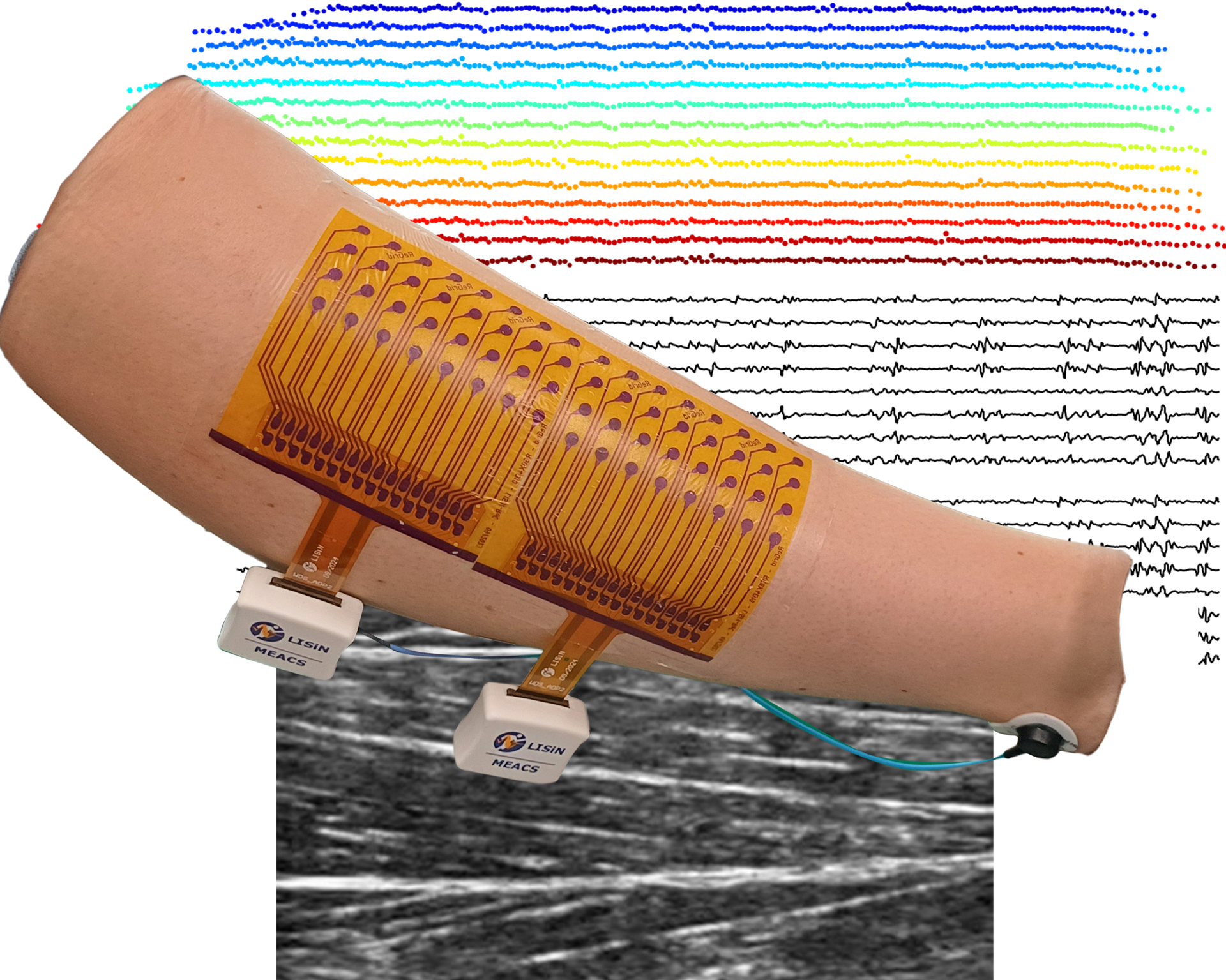
One key innovation in the HD-sEMG field is ReGrid® [1], a highly conformable and fully ultrasound transparent HD-sEMG electrode patch. ReGrid® technology adapts to curved body shapes in both static and dynamic conditions, enabling HD-sEMG acquisitions in new and challenging conditions. ReGrid® ensures electrode-skin contact quality comparable to that of standard, gel-based, HD-sEMG grids. The high quality of the collected signals enables the extraction of both global sEMG and single motor unit variables.
The MEACS system, combined with ReGrid® detection system is significantly advancing research in neuromechanics by enabling concurrent acquisitions of HD-sEMG signals and ultrasound images.
As an example, in studies MEACS was used in combination with ultrasonography to correlate mechanical and electrophysiological motor unit properties, shedding light on neuromechanical coupling between these variables [2-4]. Altogether, ReC Bioengieering supports a robust and sophisticated technological approach to neuromechanics.
Bibliography from the LISiN-ReC group
[1] G. L. Cerone, T. Vieira, M. Gazzoni, and A. Botter, “ReGrid: a highly conformable and ultrasound transparent patch for HD-sEMG detection,” submitted to the IEEE. Preprint available at the following DOI link: https://doi.org/10.36227/techrxiv.175099860.02131635/v1.
[2] E. Martinez-Valdes et al., “Modulations in motor unit discharge are related to changes in fascicle length during isometric contractions,” J. Appl. Physiol., vol. 133, no. 5, pp. 1136–1148, Oct. 2022.
[3] M. Carbonaro, K. M. Meiburger, S. Seoni, E. F. Hodson‑Tole, T. Vieira, and A. Botter, “Physical and electrophysiological motor unit characteristics are revealed with simultaneous high-density electromyography and ultrafast ultrasound imaging,” Sci. Rep., vol. 12, no. 1, p. 8855, 2022, doi: https://doi.org/10.1038/s41598-022-12999-4.
[4] R. Rohlén, M. Carbonaro, G. L. Cerone, K. M. Meiburger, A. Botter, and C. Gronlund, “Spatially repeatable components from ultrafast ultrasound are associated with motor unit activity in human isometric contractions,” J. Neural Eng., vol. 20, no. 046016, 2023, doi: https://doi.org/10.1101/2023.04.17.537211.